Retinal imaging markers of accumulated blood pressure burden
Novel retinal vascular imaging with potential for non-invasive assessment of the microvascular sequelae of hypertension.

An image of a portion of a woman's face, just the portion with the eyes
We’re developing a non-invasive assessment to identify abnormalities in the retinal microvasculature.
The challenge
Retinal markers could potentially serve as an integral summation of the blood pressure burden a patient has been exposed to at any given time. They could provide a simple, non-invasive and inexpensive test for accurate prediction of cardiovascular risk.
Changes in retinal markers over time may also serve as an indicator of the effectiveness of antihypertensive treatment.
Our response
Our project aims to identify novel retinal imaging markers that correlate closely with blood pressure measurements and other signs of hypertensive target organ damage in high-risk patients.
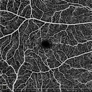
An OCT-A image showing retinal capillaries.
An OCT-A image showing retinal capillaries.
Retinal photography and optical coherence tomography-angiography (OCT-A) are utilised to investigate the structure of the retinal nerve cell layers and vasculature.
Our VASP (Vessel Analysis Software Platform) software is used to analyse the vasculature.
The results
Preliminary results indicate an association between retinal capillary rarefaction (from OCT-A imaging) and arterial stiffness, measured by pulse wave velocity (PWV).
This ongoing project involves identifying novel retinal imaging markers to develop a non-invasive means to measure and quantify subtle variations and abnormalities in the retinal microvasculature.
The project is expected to enable the creation of a simple, non-invasive and inexpensive clinic-based test for accurate prediction of cardiovascular risk in patients.
The Australian e-Health Research Centre (AEHRC) is CSIRO's digital health research program and a joint venture between CSIRO and the Queensland Government. The AEHRC works with state and federal health agencies, clinical research groups and health businesses around Australia.