HIST – Automated quantification of white matter hyperintensity burden
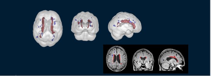
Images of brain scans showing white matter lesions
HIST provides automated image analysis for volumetric quantification of white matter hyperintensities.
Our image analysis tool can help diagnose cerebrovascular disease.
The challenge
White matter hyperintensities (WMHs) are common among older people and are associated with a high risk of cognitive decline and dementia.
However, the presence of WMHs is not usually quantified in clinical practice as it relies on tedious and time-consuming manual tracing over the image. It offers only minimal information about the severity of WMHs.
Nowadays, with increasing number of large-scale imaging studies, manual/semi-automated quantification methods are even less feasible to implement.
A robust and fully quantitative assessment tool of WMHs is desired in clinical studies and research. It would help increase understanding of cerebrovascular disease and its association with dementia.
Our response
We’ve developed an automated image analysis tool for volumetric quantification of WMHs.
This tool delivers a quantification report that offers a comprehensive inventory of WMH burden. The report includes a summary of volumetric measures, 3D visualisation of the spatial distribution as well as a reference chart comparing with the age-matched reference population.
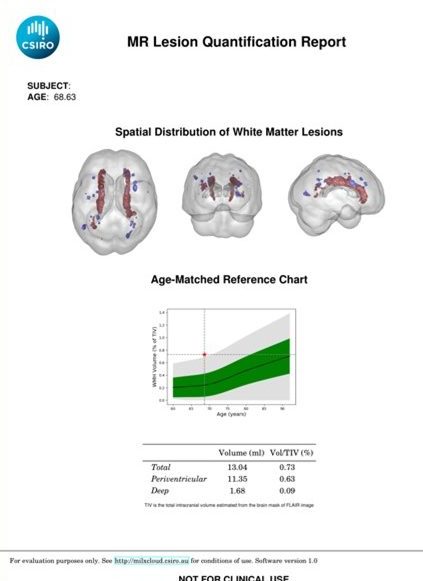
Image of an MR Lesion Quantification Report. It shows brain scans revealing the spatial distribution of white matter lesions, for a 68 year old patient, with an aged-matched reference chart.
Cerebrovascular disease is preventable once identified. HIST helps health professionals diagnose more confidently and quickly.
With this work we have also shown a high prevalence of comorbid cerebrovascular disease in patients with Alzheimer’s disease. Comorbidity of cerebrovascular disease and Aβ (peptides involved in Alzheimer’s disease) was associated with cognitive decline and neurodegeneration.
Benefits
Using the CSIRO technology, health professionals will be able to provide a timely and confident diagnosis of cerebrovascular disease. Interventions can then be put in place – the disease is preventable using evidence-based lifestyle and pharmacological approaches.
The tool also enables the detection and characterisation of longitudinal changes in WMHs. This could be used as a biomarker for disease progression and to further guide the development of treatment and prevention strategies targeting vascular risk factors.
The Australian e-Health Research Centre (AEHRC) is CSIRO's digital health research program and a joint venture between CSIRO and the Queensland Government. The AEHRC works with state and federal health agencies, clinical research groups and health businesses around Australia.